Amazing Images of a Baby Developing in the Womb: Childbirth
Swedish photographer Lennart Nilsson spent 12 years of his life taking pictures of the foetus Developing in the womb. These incredible photographs were taken with conventional cameras with macro lenses, an endoscope and scanning electron microscope. Nilsson used a magnification of hundreds of thousands and “worked” right in the womb. His first photo of the human foetus was taken in 1965. Lennart Nilsson managed to get his most precise shots with the help of a cystoscope – a medical instrument which is used to examine the inside of the urinary bladder. He attached a camera together with a tiny light to it, and took thousands of photos recording the life of the embryo in its mother’s womb.
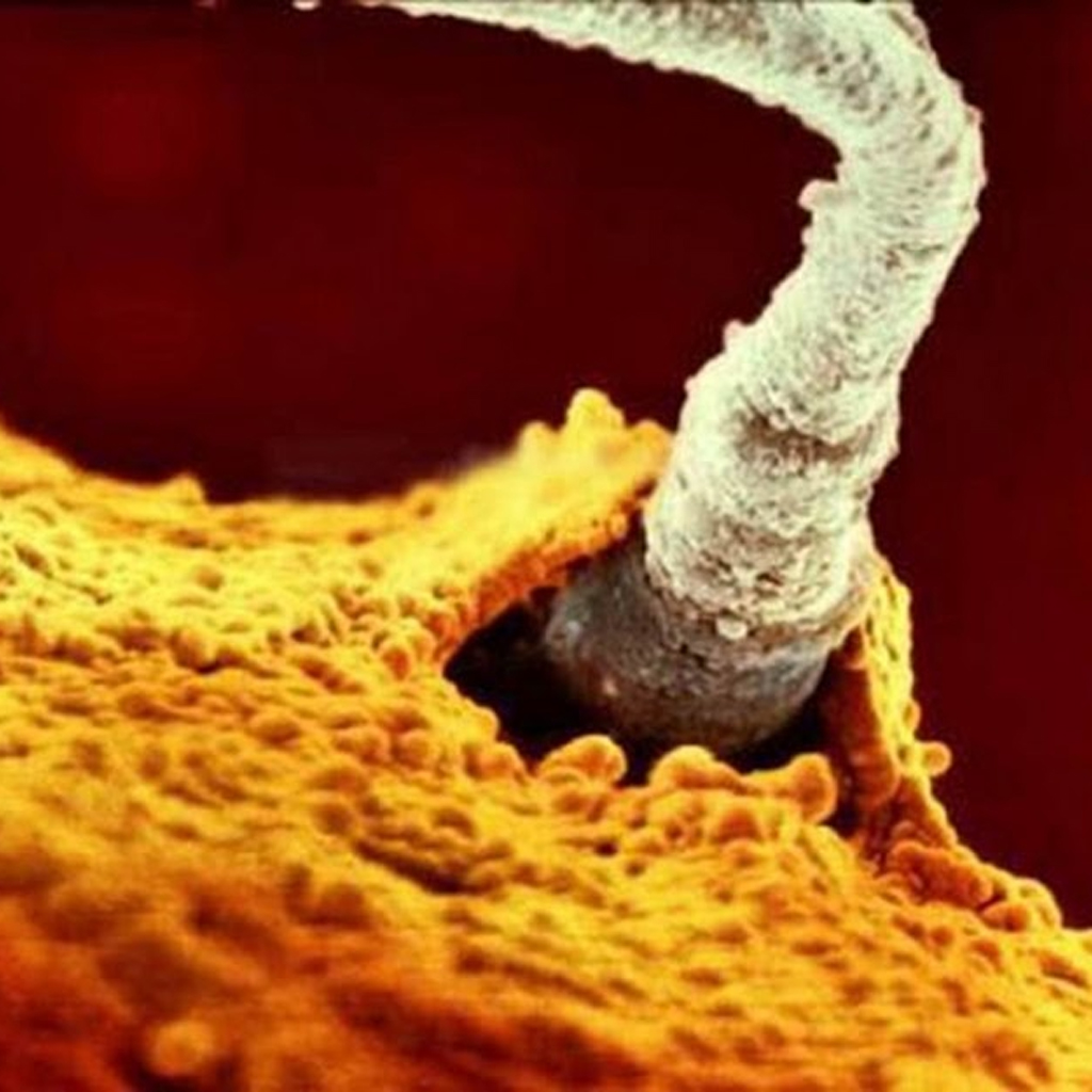
Sperm as it makes its way down the fallopian tube.
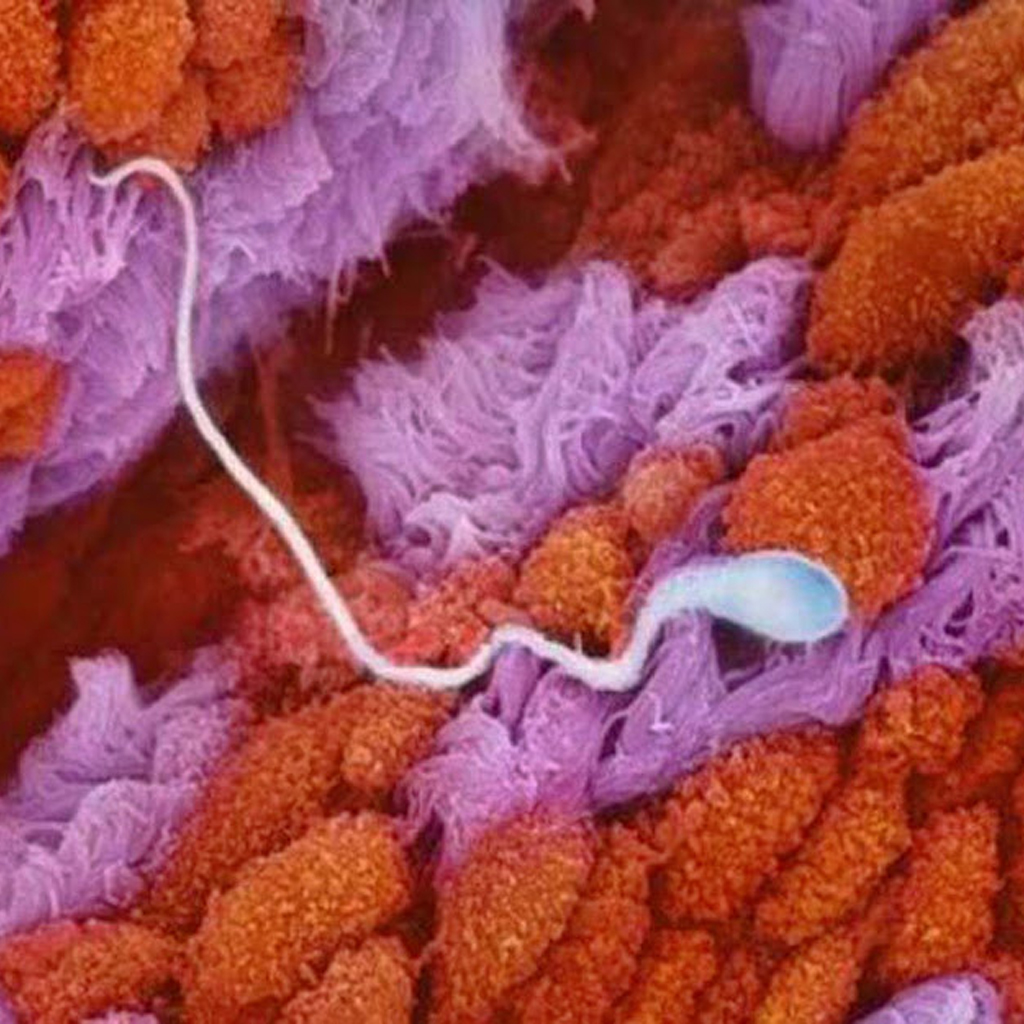
The sperm approaches the egg cell.
Amazing Images of a Baby Developing in the Womb: The Birth of a Child
Two sperms make contact with the egg cell.
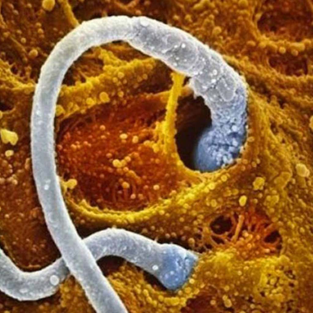
The winning sperm penetrates the surface of the egg cell.
The moment of conception as the sperm enters the egg cell and fertilizes it.

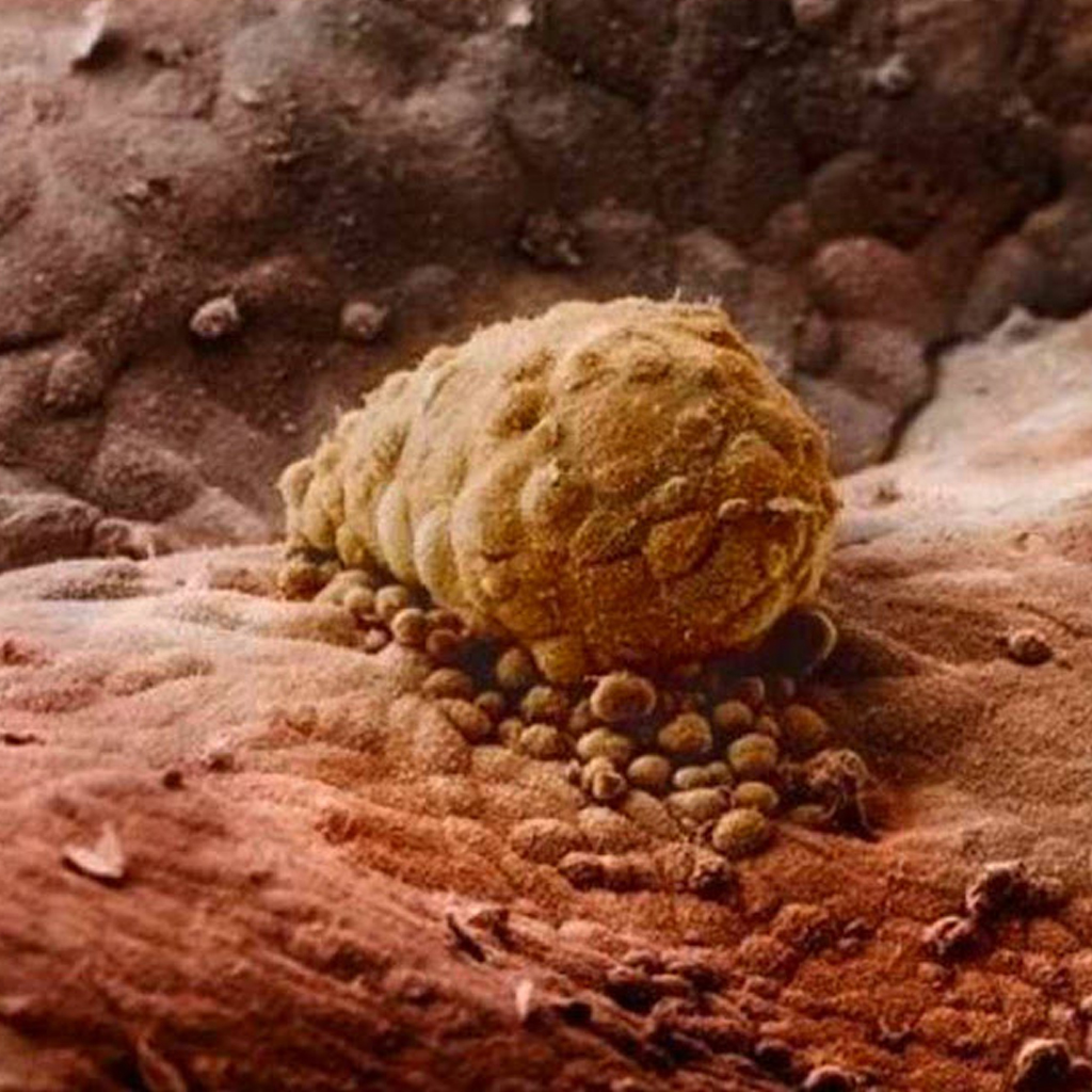
After eight days, the human embryo is attached to a wall within the womb.
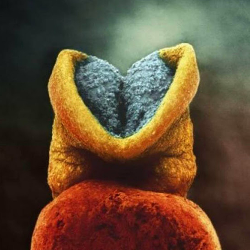
The brain starts to develop inside the human embryo.
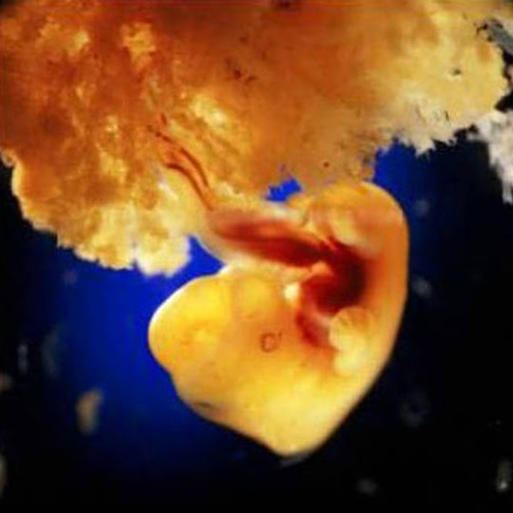
At 24 days the one-month-old embryo has a heart that started beating on the 18th day.

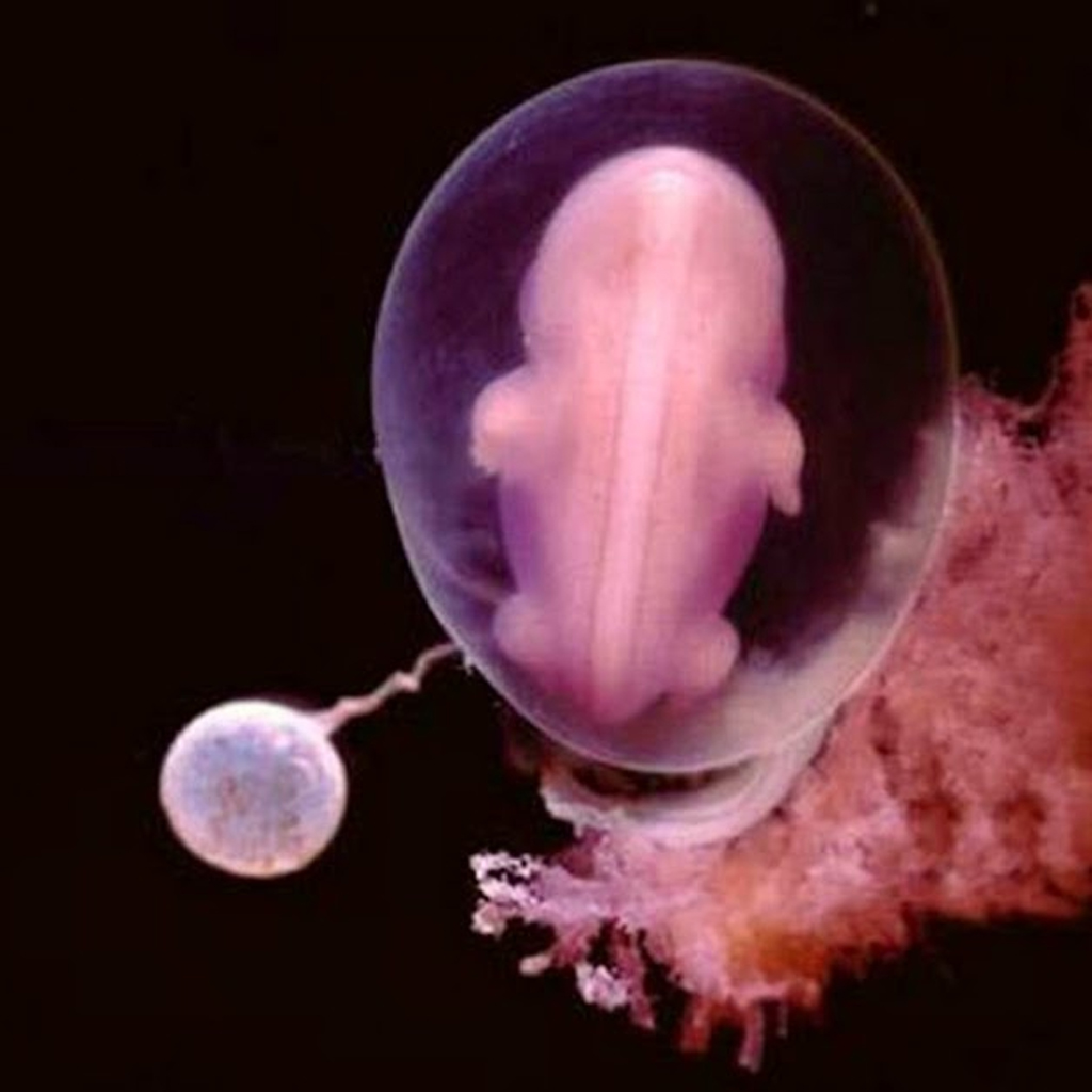
After four weeks you can start to distinguish the human shape as a skeleton starts to form.
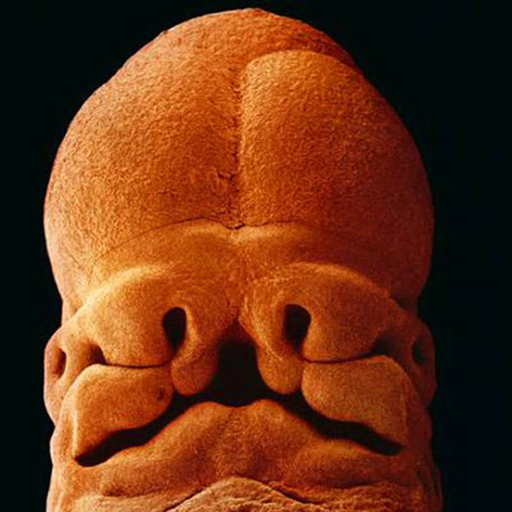
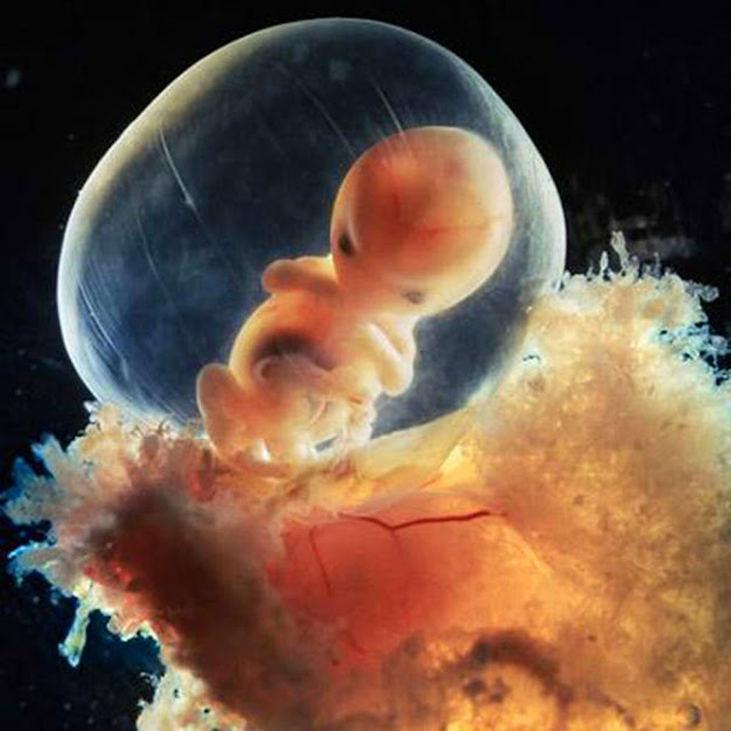
At five weeks the face begins to take shape, with holes for the eyes, nostrils and mouth. The embryo is now around 9mm in size.
After 40 days the placenta starts to be formed. This organ connects the embryo to the womb so that the developing fetus can take in nutrients, oxygen and get rid of waste via the mother’s blooᴅ supply
At eight weeks the growing embryo is well protected in its fetal sack.
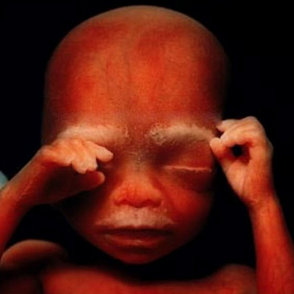
The fetus starts to use its hands to explore its body and surroundings after 16 weeks.
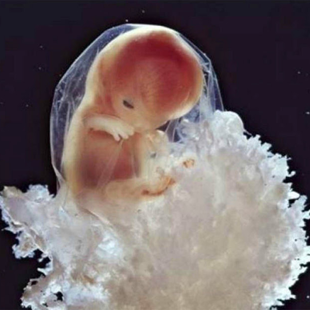
The tiny fetus is very much starting to look like a baby.
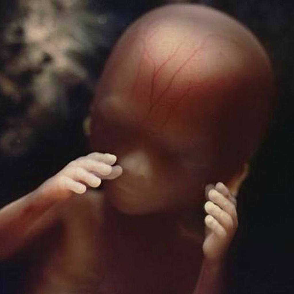
At this point the fetus’ skeleton consists of flexible cartilage while blooᴅ vessels can be seen through the thin skin.
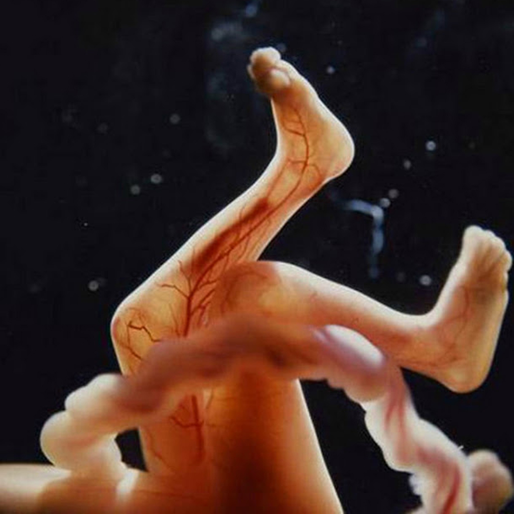
At this point the fetus’ skeleton consists of flexible cartilage while blooᴅ vessels can be seen through the thin skin.
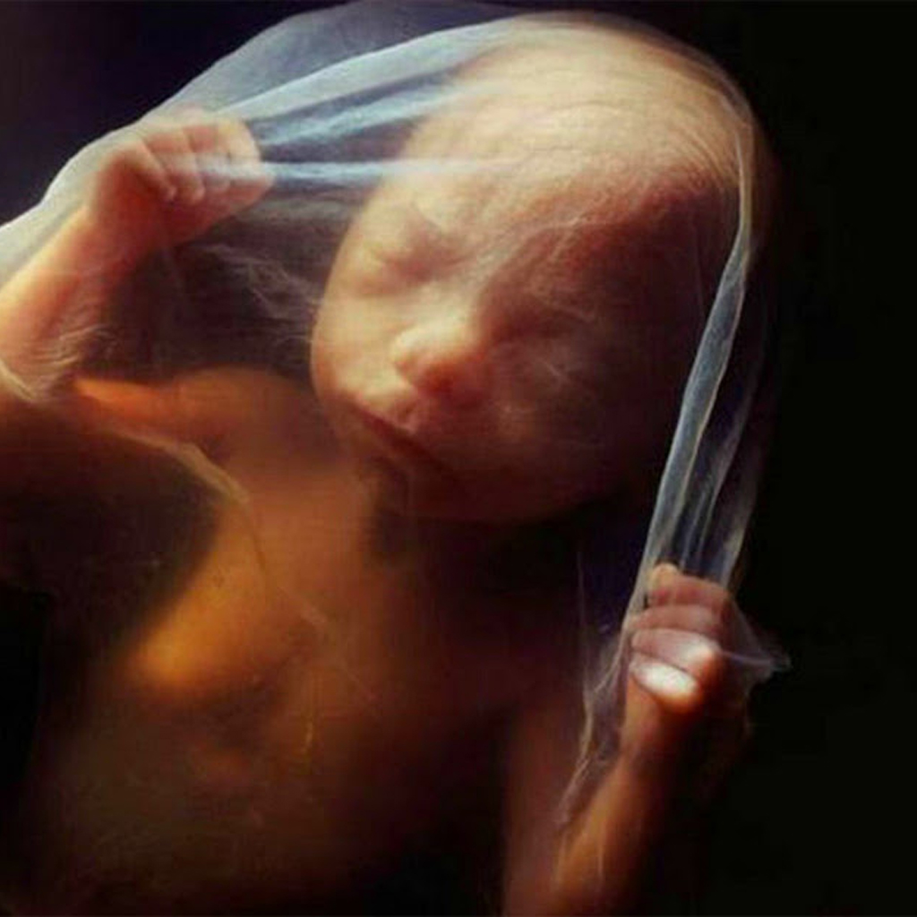
At 18 weeks the fetus is now around 18cm and can hear sounds from the outside world.

After 19 weeks the fetus has grown tiny fingernails.
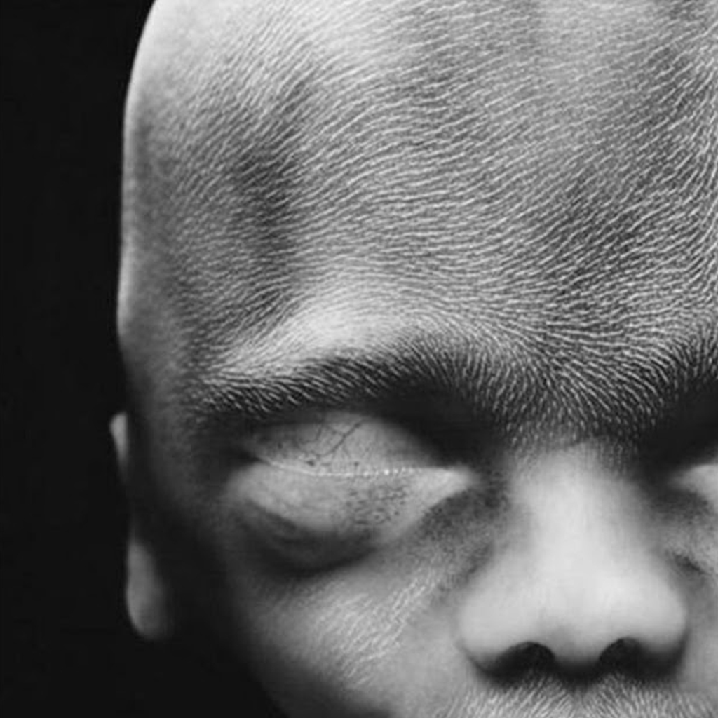
At 20 weeks wooly hair known as lanugo covers the fetus’ head. In two weeks the baby has grown an additional 2cm.
Here is the fetus after 26 weeks.

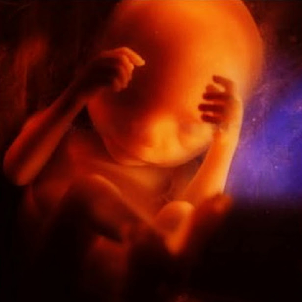
After six months the fetus is preparing to leave the womb. It turns upside down because it will be easier to get out this way.
Four weeks before the baby is due to enter the world.
Hits: 3